A fetal pig is a great choice for dissection because the size of the organs makes them easy to find and identify.
It is also a very exciting dissection because, like sheep and their organs, the internal anatomy is similar to humans! It is fascinating to see how all the organs fit and work together. Use this guide to help you dissect a preserved fetal pig, or just look at the labeled pictures to get an idea of what the organs look like. If you do the dissection yourself, you will need dissection pans and dissection tools, or buy our complete Fetal Pig Dissection Kit.
External Anatomy
1. Most of the pig’s external features are familiar to you – ears, nose, eyes, etc. On the belly you will see the umbilical cord which connected the fetal pig to its mother’s placenta. On either side of the umbilical cord, you may see mammary papillae, little nipples that will turn into teats in female pigs.
2. Determine if your specimen is male or female by looking closely at the umbilical cord area. If the pig is male, it will have a small urogenital opening immediately behind the umbilical cord. If the pig is female, the urogenital opening will be just behind the anus under the pig’s tail. Only the female has two openings beneath the tail.
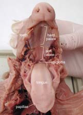
Oral Cavity
|
| Click image for full-size pdf |
1. Using your dissecting scissors, cut through the jawbones at the corner of the pig’s mouth. Cut far enough so that the bottom half of the jaw can almost touch the pig’s chest.
2. Open the mouth as far as you can. Use the labeled picture to identify the feathery papillae (taste buds) on the edges of the tongue, the ridged hard palate in the roof of the mouth with the smooth soft palate behind it, the sharp teeth near the front of the mouth, and the epiglottis, which covers the opening of the trachea (windpipe) so food cannot enter.
Body Cavity Incisions
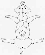
1. Tie a string around one of the pig’s forelegs. Pass the string under your dissecting pan and tie it to the other foreleg. Stretch the string tightly so that it will hold the pig’s legs apart. Repeat with the back legs.
 |
| Click image for full-size pdf |
2. Use your fingers to probe the chest area of the pig. You should be able to feel the hard sternum (breastbone) and the tiny ridges of the ribcage. Keep moving down until you feel the bottom edge of the rib cage; this is where the diaphragm separates the thoracic cavity from the abdominal cavity. This point is marked with an X in the illustration.
3. Make an upside-down V incision starting at X, as you see in the illustration. Start the cut with a plastic scalpel, then continue it with dissecting scissors. You want to cut through the skin and the muscle, but if you cut too deep you will damage the internal organs. Use forceps to hold the tissue away from the organs as you cut. Carry the incision all the way to the pan.
4. Start the second incision at X and carry it straight down almost to the umbilical cord. Cut a semi-circle around the umbilical cord on each side. Make the third incision just above the hind legs and carry it all the way down to the pan.
5. Now lift up the flaps of skin and peel them back so they lay flat on the pan. There may be some connective tissue or membranes attaching the muscles to the underlying organs. Cut carefully through this so you can lift the flaps back. The abdominal cavity is now exposed.
6. Beginning at X again, make the fourth incision up through the chest. Use the scissors to cut through the rib cage and the sternum. When you reach the midpoint between the forelegs, make another incision down to the pan. Go back to the diaphragm area and use a scalpel to cut the wall of the body cavity away from the diaphragm. The diaphragm should remain intact, but now the rib cage can be pulled back and pinned to the pan, exposing the thoracic cavity.
7. Make the last two incisions to expose the neck area.
Abdominal Cavity
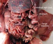
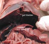
1. The largest organ in the abdominal cavity is by far the liver, just below the diaphragm (the flap of muscle separating the abdominal from the thoracic cavity). Notice the umbilical vein connecting the umbilical cord with the liver. Cut this vein so you can lay the umbilical cord back between the pig’s hind legs.
2. Use the labeled pictures to find the following organs:
 |
| Click for full-size pdf |
 |
| Click for full-size pdf |
3. If your specimen is a male, you will find long brown tubes on either side of the folded-back umbilical cord. At the end of these tubes are the rounded testes. A whitish sack attached to the testis is the epididymis, which stores sperm cells. If your specimen is female, you will find the ovaries at the base of the umbilical cord/urinary bladder. Directly below the ovaries, you will see a flap of tissue called the horns of the uterus; this part leads to the main body of the uterus.
Thoracic Cavity
1. The thoracic cavity is protected by the rib cage and contains the lungs and heart. Use the labeled picture to find the following organs:
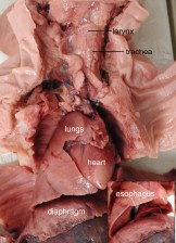 |
| Click for full-size pdf |
To study the pig in more detail, go to this Virtual Pig Dissection. It covers all the body systems and includes quizzes to test your knowledge!
See our other free dissection guides with photos and printable PDFs.
What other users say:
Absolutely Fantastic!
My students thoroughly enjoy completing the dissection projects. I highly recommend these specimens for anyone interested in learning more about the anatomy of specific organisms. -Sandi
Welcome! Read other Biology / Life Science articles or explore our the rest of the Resource Center which consists of hundreds of free science articles!